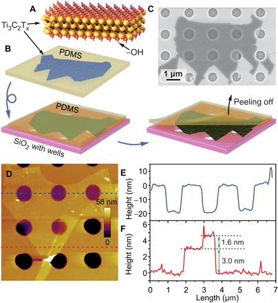
Fig. 1. Preparation of MXene membranes.

(A) Structure of a Ti3C2Tx monolayer. Yellow spheres, Ti; black spheres, C; red spheres, O; gray spheres, H. (B) Scheme of the polydimethylsiloxane (PDMS)–assisted transfer of MXene flake on a Si/SiO2 substrate with prefabricated microwells. See text for details. (C) SEM image of a Ti3C2Tx flake covering an array of circular wells in a Si/SiO2 substrate with diameters of 0.82 μm. (D) Noncontact AFM image of Ti3C2Tx membranes. (E and F) Height profiles along the dashed blue (E) and red (F) lines shown in (D).
