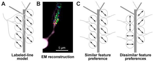
Figure 1. Models for convergence of retinal axons in dLGN thalamus.
A. Labeled-line model: all or most retinal boutons contacting a dLGN neuron proximal dendrite (dark gray) arise from a single RGC axon (light gray).
B. Electron microscopy reconstruction demonstrates boutons from multiple RGC axons (different colors) contacting the same dLGN neuron dendritic domain. Adapted from Morgan et al., 2016.
C. Different axons contacting the same dLGN neuron could exhibit the same visual feature preference (left; arrows indicate common preference for axis of motion) or random preferences (right).

