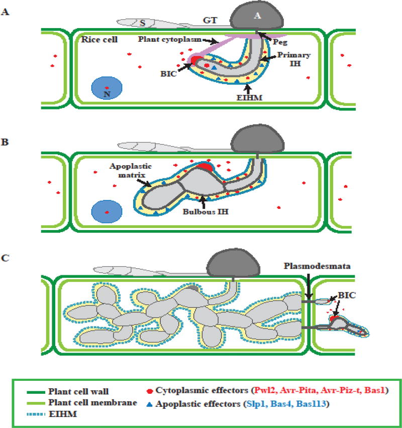
Figure 3.
BIC development and effector secretion during biotrophic growth. These illustrations represent the growth of the invasive hyphae (IH) from 22–40 hour post inoculation inside rice cells. (A) BIC develops at the tip of the primary filamentous invasive hyphae (IH). (B) BIC localizes at a subapical position in the first bulbous IH, and remains behind as the fungus grow in the rice cell. (C) New BICs are formed in the IH tip in the adjacent cells. Cytoplasmic effectors (red circles), including Pwl2, Avr-Pita, Avr-Piz-t and Bas1, accumulate at the BIC and are secreted into the host cytoplasm. In contract, apoplastic effectors (blue triangles), including Slp1, Bas4 and Bas113 are secreted into the apoplastic matrix between EIHM and fungal cell wall. Abbreviations: S, Spores; GT, Germ tube; A, Appressorium; N, Nucleus; BIC, Biotropic interface complex; IH, Invasive hyphae; EIHM, Extrainvasive invasive hyphae membrane

