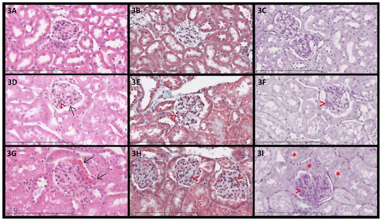
Figure 3. Histopathology of the kidney.
(original magnification 400x). 3A, 3D and 3G: H & E stained sections; 3B, 3E and 3H: Masson trichrome stained sections; 3C, 3F and 3I: Periodic acid-Schiff stained sections. 3A–C: Glomeruli from Hb AA mouse with normal cellularity and unremarkable tubules. 3D–F: Glomeruli from Hb AS mouse with mild focal segmental mesangial hypercellularity (>) and segmental congestion of glomerular capillary loops (→). 3G–I: Glomeruli from Hb SS mouse with global diffuse mesangial hypercellularity (>) and segmental congestion of glomerular capillary loops (→). 3I: Hb SS mouse with tubular and parietal epithelial deposition of cytoplasmic hemosiderin pigment (*).

