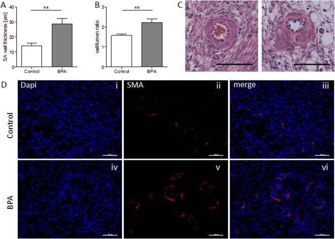
Figure 5.
Impaired uSA remodeling of BPA-treated mice at gd10. uSA thickness (A) and wall-to-lumen ratios (B) from C57BL/6J control (n = 7) and BPA-treated C57BL/6J mice (n = 5) were calculated by measuring wall and lumen diameters of 3–8 uSAs per mouse. Results are presented as means ± S.E.M. and analyzed by using Mann-Whitney-U-test (**P < 0.01). (C) Representative images of uSAs from H/E-stained implantations of C57BL/6J (i) and BPA-treated C57BL/6J (ii) are shown (scale bars = 100 µm). (D) Representative smooth muscle actin immunfluorescence staining images of SAs from implantations of BL/6J (i-iii) and BPA-treated BL/6J (iv-vi) are shown (scale bars = 100 µm). uSA, uterine spiral artery; gd, gestation day.

