Fig. 1.
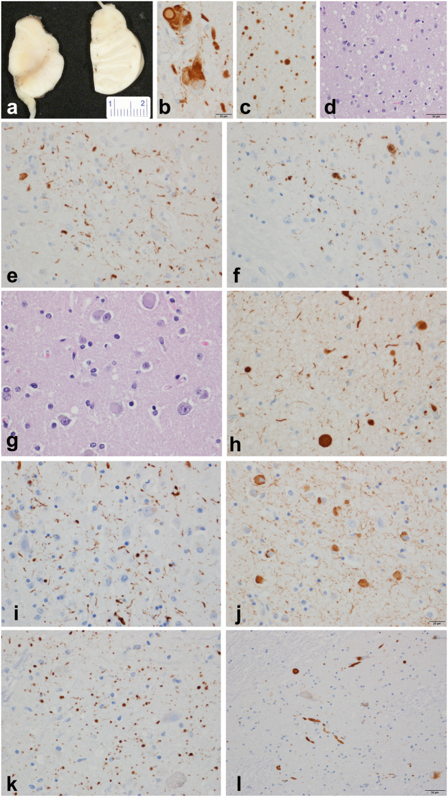
SNCA genomic triplication neuropathology. a Gross pathology of midbrain and pons with loss of neuromelanin pigment in both; b alpha-synuclein immunohistochemistry of locus ceruleus with Lewy bodies and bizarre neuronal inclusions; c Numerous Lewy dots in ventral tegmental region of midbrain; d spongiform change in neocortex in temporal and limbic lobes; e CA2 sector of hippocampus with Lewy neurites; f CA2 sector of hippocampus with tau in subset of Lewy neurites; g, h cortical Lewy bodies in temporal neocortex; cortical Lewy bodies and Lewy neurites in temporal neocortex; i hippocampal CA2 neurites; j amygdala Lewy bodies and neurites; k ventral tegmental area Lewy neurites (“Lewy dots”); l substantia nigra pars compacta Lewy bodies.alpha-synuclein immunohistochemistry (b, c, e, h), phospho alpha-synuclein (i, j, k, l), tau immunohistochemistry (f), hematoxylin and eosin stain (d, g). Bar in b = 20 μm (applies to c, e, f, g, h, i, j, and k); bar in d and l = 50 μm; measure bar in a is in mm

