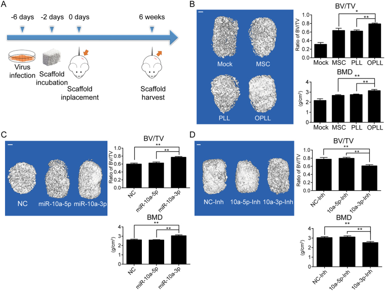
Figure 6.
MiR-10a-3p promotes the ossification of OPLL in vivo. (A) A scheme of the procedure in heterotopic bone formation assay. (B) Reconstructed 3D micro-CT images of the tissue-engineered bone constructs from different type of cells seeded (n = 6). The ratio of bone volume to tissue volume (BV/TV) and bone mineral density (BMD) of cultured bone constructs were shown in the right panel. *P < 0.05, **P < 0.01, t-test. (C) Reconstructed 3D micro-CT images of the tissue-engineered bone constructs that contains miR-10a-3p, miR-10a-5p or negative control stably expressed PLL cells (n = 6). The ratio of bone volume to tissue volume (BV/TV) and bone mineral density (BMD) of cultured bone constructs were shown in the right panel. **P < 0.01, t-test. (D) Reconstructed 3D micro-CT images of the tissue-engineered bone constructs that contains miR-10a-3p, miR-10a-5p or negative control stably inhibited OPLL cells (n = 6). The ratio of bone volume to tissue volume (BV/TV) and bone mineral density (BMD) of cultured bone constructs were shown in the right panel. **P < 0.01, t-test. The error bars represents ±S.D.

