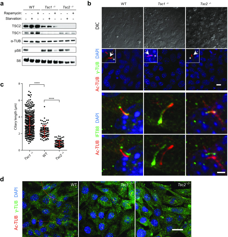
Fig. 1.

Primary cilia are elongated in Tsc1−/− but shortened in Tsc2−/− cells. a SDS-PAGE and WB analyses of cell lysates from WT, Tsc1−/− and Tsc2−/− MEFs cultured in normal (-starvation) or starvation medium (0.5% FBS) for 48 h, using antibodies against TSC1, TSC2, S6 and phospho-S6 (pS6) as indicated, with antibody against α-tubulin (α-TUB) as loading control. Note the presence of pS6 in serum-starved Tsc1−/− and Tsc2−/− cells. b IFM analysis of primary cilia in WT, Tsc1−/− and Tsc2−/− MEFs cultured in starvation medium (0.5% FBS) for 48 h, to induce ciliogenesis. Cilia (arrows) were labeled with anti-acetylated α-tubulin (Ac-TUB) and anti-IFT88 antibodies and the ciliary base/centrosomes (asterisks) were labeled with anti-γ-tubulin (γ-TUB) antibody. Nuclei were visualized with differential interference contrast (DIC) microscopy or DAPI staining. Lower panels show merged and shifted overlays of Ac-TUB- and IFT88-labeled cilia. Scale bar: 2 µM. c Quantification of cilia lengths for experiment shown in b. Three hundred cilia (Ac-TUB) from three independent experiments were used for quantification. d IFM analysis of WT, Tsc1−/− and Tsc2−/− MEFs labeled with DAPI, Ac-TUB and IFT88 for cell size comparison. Nuclei were visualized with DAPI staining. Scale bar: 5 µM
