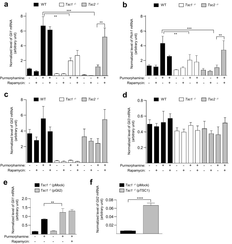
Fig. 4.
Loss of Tsc1 or Tsc2 is associated with impaired HH signaling. a–d WT, Tsc1−/− and Tsc2−/− MEFs were starved (0.5% FBS) for 48 h to induce cilia formation in the presence or absence of rapamycin. Purmorphamine (5 µM) was administered to the medium for the final 24 h to stimulate HH signaling before mRNA isolation and qPCR analysis. Expression profiles of the target genes were normalized to the amount of endogenous Tbp mRNA. Normalized expression profiles of a Gli1, b Ptch1. Error bars represent SEM (n = 5). Normalized expression profiles of c Gli2 and d Gli3. e Normalized expression profiles of Gli1 in Tsc1−/− MEFs transfected with 2 µg plasmid (pMock (empty vector) or pGli2) and cultured in starvation medium (0.5% FBS) for 48 h (24 h after transfection) with purmorphamine administration (for the last 24 h), as indicated. Error bars represent SEM (n = 3). The reduced signal in e compared to a might be due to transfection (pMock included) of the cells. f Normalized expression profiles of Gli2 in Tsc1−/− MEFs transfected with 1 µg plasmid (pMock or pTSC1) and starved (0.5% serum) for 48 h (24 h after transfection). Error bars represent SEM (n = 3)

