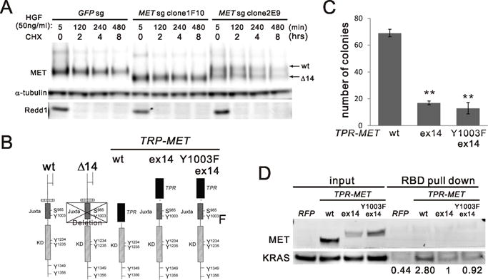
Figure 3.

A) MET protein half-life. AALE GFPsg cells or METsg single clone 1F10 and 2E9 were treated with cycloheximide (CHX) for indicated time after 5 min of HGF stimulation and washout. Cell lysates were immunoblotted with MET antibody and α-tubulin antibody and Redd1 antibody for CHX control. B) a schematic panel of MET protein domains in wild type (wt) and mutants. Exon 14 skipping (Δ14), exon 14 (ex14). C) Comparing AALE cells with stable ectopic expression of V5 tagged TPR-MET wt (wt) and mutants including wt exon14 insertion (ex14) and Y1003F exon14 insertion (Y1003F ex14). Anchorage-independent growth was assessed as described in (Fig. 2A). **p<0.01. D) RAS-binding domain (RBD) pull down assay comparing RAS activation in AALE cells described in (C).
