Figure 6.
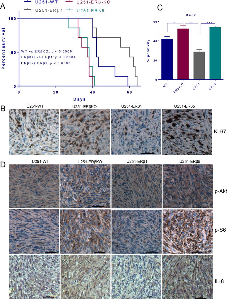
Differential effects of ERβ1 and ERβ5 isoforms on the progression of GBM in vivo. A. Athymic nude mice were implanted with U251-WT or U251-ERβ-KO or U251 ERβ1 or U251 ERβ5 cells orthotopically into the right cerebrum. Survival of the mice was plotted using Kaplan-Meier curve. B. Mouse brains collected from the WT, KO, ERβ1 and ERβ5 groups were fixed in formalin and processed for immunohistochemical staining for Ki-67. C. The number of Ki-67 positive cells from five different images were counted and plotted as histogram. D. Tumor sections were subjected to immunohistochemical staining for the detection of p-Akt, p-S6, and IL-8. Data are represented as mean ± SE. * p<0.05. ** p<0.01.*** p<0.001.

