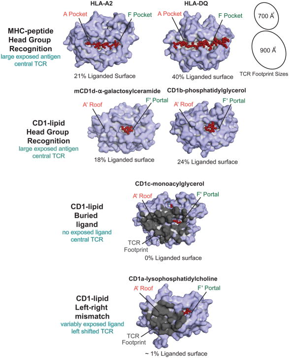
Figure 2. The Exposed Surfaces of MHC-peptide and CD1-Lipid complexes.
Peptides are broadly exposed across most of the MHC I platform and across the entire MHC II platform. Idealized TCR footprints (black ovals, shown to scale) are difficult to place on MHC platforms without contacting peptide. In contrast, the A′ roof provides a large, ligand-free landing surface for TCRs on CD1 proteins. Color-coding of exposed antigen surfaces (red) shows that protruding ligands are more variable and asymmetrically positioned on CD1 platforms as compared to peptides on MHC platforms. The buried ligand mechanism describes bound lipids that fail to protrude to the surface. In the left-right mismatch mechanism, TCRs have left shifted footprints (dark grey) and the ligand (red) protrudes toward the right side of the platform.

