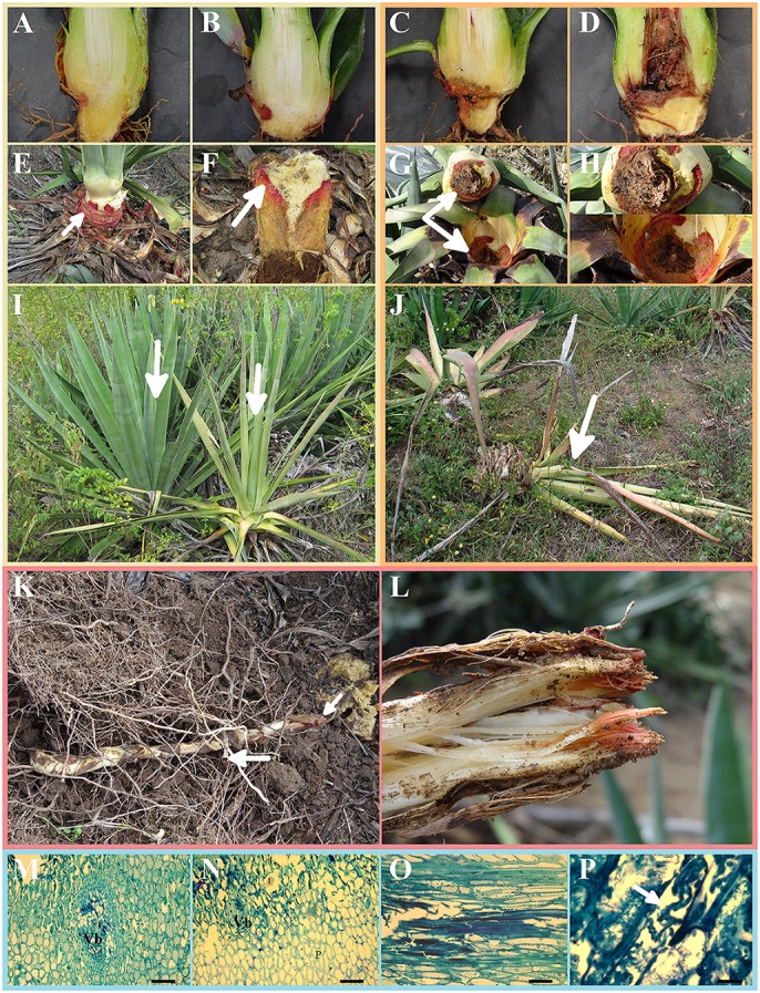
Figure 2.

Symptoms of bole rot disease in healthy sisal bulbils grown under greenhouse conditions and adult plants in the field (I–L) and histopathology of the infected boles of adult plants (M–P). (A–D) Gross longitudinal sections of an artificially infected sisal bulbil showing the internal lesion of the aerial stem. (E) External view of an infected aerial stem showing the typical reddish color due to the fungal infection (white arrow). (F) Detailed view of the internal infected tissues of the aerial stem (white arrow). (G,H) Gross transversal sections in distinct magnifications showing the necrotic lesions of the aerial stem. (I) Healthy adult plant (left white arrow) beside a symptomatic adult plant (right white arrow) in the field. (J) Dead adult plant due to fungal infection (white arrow). (K) An external view of a symptomatic rhizome (subterraneous stem) of an adult plant in the field. (L) Gross longitudinal section of a rhizome of an adult plant in the field. (M,N) Histological transverse sections of an infected aerial stem of an adult plant, showing a degraded vascular bundle, parenchymatic cells and idioblast. VB, vascular bundle; (P) parenchyma; I, idioblast; (O,P) histological longitudinal sections of an infected aerial stem of an adult plant at different magnifications, showing fungal hyphae in intercellular spaces and inside parenchymatic cells (white arrow).
