Fig. 1.
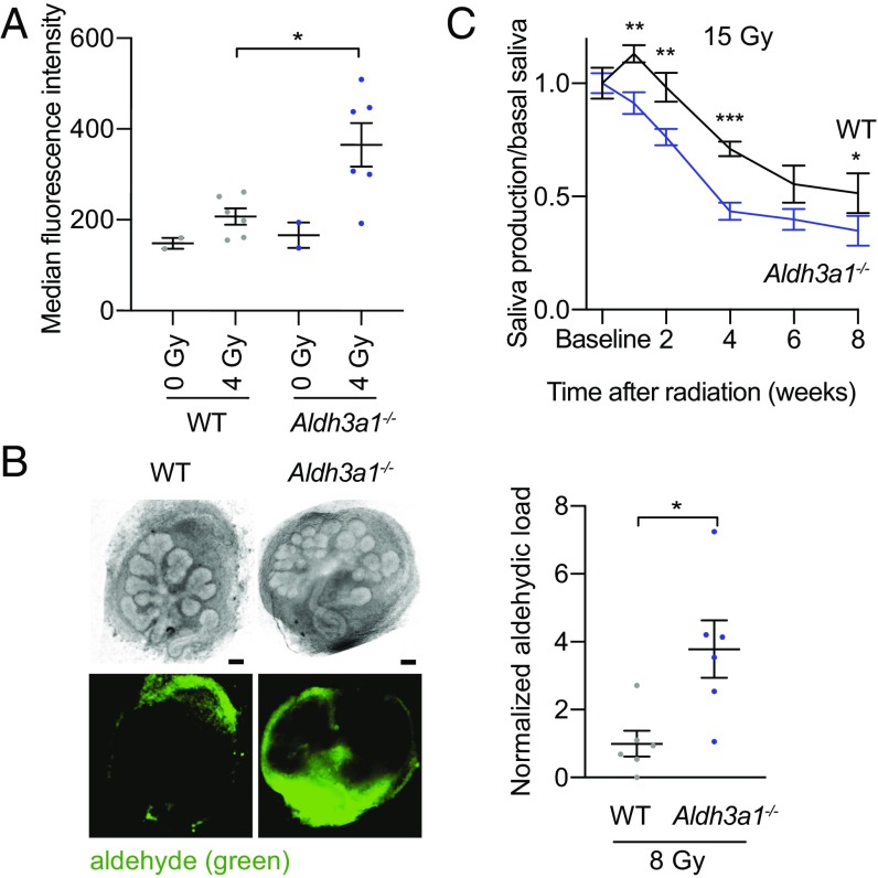
Loss of ALDH3A1 increases aldehyde accumulation in SSPCs and accelerates hyposalivation after radiation. (A) Aldehyde levels in dissociated WT and Aldh3a1−/− murine salispheres 2 h after IR, measured as median fluorescence intensity of DarkZone dye by FACS (n = 2–6; bars indicate SEM; *P < 0.05). The experiment was repeated in SI Appendix, Fig. S1A. (B, Left) Representative images of WT (Left) and Aldh3a1−/− (Right) E13.5 mouse SMGs after 24 h in culture, treated with DarkZone dye, 3 h after IR, in brightfield (Upper Row) and with a florescence filter (Lower Row). (Scale bars: 50 μm.) (Right) Quantification of DarkZone dye fluorescence intensity of embryonic SMGs, normalized to WT (n = 6; bars indicate SEM; *P < 0.05). (C) Pilocarpine-induced saliva production collected in female C57BL/6J WT and Aldh3a1−/− mice at baseline and 1, 2, 4, 6, and 8 wk after 15-Gy IR (single dose) (n = 8–11; bars indicate SEM; *P < 0.05; **P < 0.01; ***P < 0.001).

