Fig. 2.
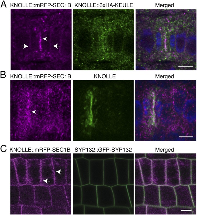
Subcellular localization of KNOLLE::mRFP-SEC1B. Immunofluorescence (A and B) and live-imaging (C) of KNOLLE::mRFP-SEC1B in seedling roots: (A) KNOLLE::mRFP-SEC1B (magenta) and KNOLLE::6xHA-KEULE (green) labeled with anti-HA antibody in fixed seedling root. (B) KNOLLE::mRFP-SEC1B (magenta) and KNOLLE (green) labeled with anti-KNOLLE antiserum in fixed seedling root. (C) Live imaging of KNOLLE::mRFP-SEC1B (magenta) and SYP132::GFP-SYP132 (green) in seedling roots. Note that mRFP-SEC1B locates at the cell division plane (arrowheads in A and B) and plasma membrane (arrows in A and C) (see also SI Appendix, Fig. S3A). Note also that the punctate signal of mRFP-SEC1B becomes more prominent after fixation (compare also Right and Left panels in SI Appendix, Fig. S3A). DAPI was used for staining nuclei (blue). (Scale bars: 10 µm.)

