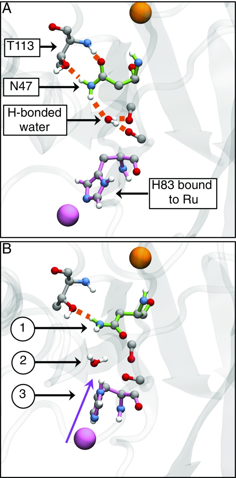
Fig. 4.
Representative WT configurations from KC-RPMD simulations illustrating the hydrogen-bonding network in the pocket surrounding residue 47. (A) The reactant basin exhibits a hydrogen-bond network between N47, the neighboring T113, a water molecule present in the pocket between N47 and H83, and nearby backbone oxygens. (B) The dividing surface is characterized by changes that include (1) breaking of the hydrogen bonds around N47, (2) displacement of the water molecule from the pocket, and (3) compression of H83 into the pocket. Hydrogen bonds are indicated by orange-dashed lines, the Cu center is in orange, residue 47 is highlighted in green, and the Ru center and H83 are highlighted in pink.

