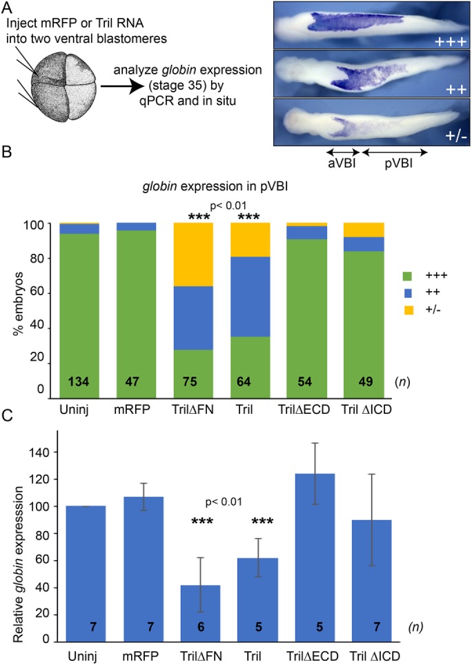
FIGURE 2:

Overexpression of Tril or Tril∆FN inhibits blood formation. (A, B) RNA encoding mRFP or wild-type or deletion mutant forms of Tril was injected into both ventral cells of four-cell embryos, and expression of hba3.L was analyzed by WMISH at stage 34 in three experiments. hba3.L staining in the posterior VBI (pVBI) was scored as absent or very weak (+/–), weak (++), or strong (+++), as illustrated. aVBI, anterior VBI. n represents the number of embryos analyzed in each group, pooled from three independent biological replicates. (C) RNA encoding mRFP or wild-type or deletion mutant forms of Tril was injected into both ventral cells of four-cell embryos and expression of hba3.L was analyzed in 15 pooled embryos from each group by qPCR at stage 34. Mean ± SD are shown. n is the number of biological replicates, indicated on each column. ***p < 0.01 by two-tailed t test.
