FIGURE 10:
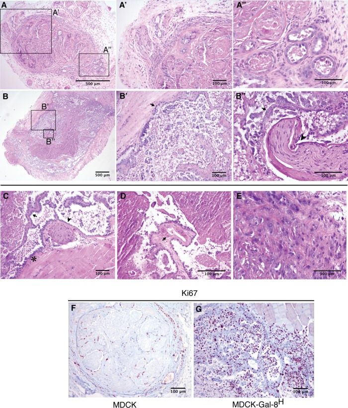
Histology and proliferation activity of MDCK- and MDCK-Gal-8H-generated tumors. Tumors generated in NSG mice subcutaneously injected with 106 cells were analyzed after 10 wk. (A–E) MDCK tumor HE staining. (A) A well-defined nodule generated by MDCK cells showing an area with small and medium vascular structures in the peripheral pseudocapsule (enlarged in A′), and a region enriched in tubules formed by cuboidal cells of differentiated appearance, filled with cellular detritus and separated by ECM and interstitial cells (enlarged in A″). (B–E) MDCK-Gal-8H tumor HE staining. (B) A tumor generated by MDCK-Gal-8H cells is labeled at regions showing an invasive behavior revealed by muscle infiltration (enlarged in B′, arrow) and perineural invasion (enlarged in B″, arrowhead) with abundant micropapillae differentiation (B″, arrow). (C–E) Selected areas of other MDCK-Gal-8H-generated tumors showing malignant features: (C) muscle infiltration (asterisk), papillae differentiation (arrow), and perineural invasion (arrowhead). (D) A papillae with a vascular structure (arrow). See higher magnifications of B, B″, C, and D in Supplemental Figure 1. (E) Undifferentiated cells with categorical tumoral appearance. (F, G) Immunohistochemistry of cell proliferation marker Ki67 shows a low percentage of cell proliferation (<15%) in MDCK nodules contrasting with the high index of cell proliferation (60–70%) in the MDCK-Gal-8H tumor.

