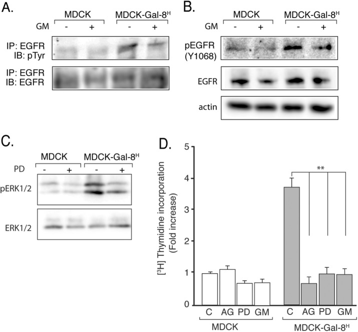
FIGURE 2:
MMP-mediated transactivation of EGFR drives the higher proliferation of Gal-8–overexpressing MDCK-Gal-8H cells. (A) Immunoprecipitated (IP) EGFR analyzed by immunoblot (IB) for tyrosine-phosphorylation (p-Tyr, top blot) and total mass (EGFR, bottom blot); (B) Immunoblot of EGFR for Tyr1068 phosphorylation (p-EGFR1068; top blot) and total mass (EGFR; middle blot) and actin in 50 μg of proteins from cell extracts of MDCK-Gal-8H and MDCK cells incubated in the absence or presence of MMP inhibitor GM6001 (GM; 2.5 μM) for 4 h. MDCK-Gal-8H cells show higher activation but similar expression of EGFR compared with MDCK cells, and their EGFR activity decreased after treatment with GM. (C) Immunoblot analysis of phospho-ERK1/2 and total mass of ERK1/2 in cell extracts (50 μg) shows higher activity of ERK1/2 in MDCK-Gal-8H cells compared with MDCK cells, which is reduced by the MEK inhibitor PD98059 (PD), indicating activation of the EGFR/RAS/RAF/MEK/ERK pathway. (D) Inhibitors of EGFR tyrosine-kinase (0.1 μM AG1478; AG), MEK (25 μM PD98059; PD), and MMP (2.5 μM GM6001; GM) all abolish the increased proliferation of MDCK-Gal-8H cells (n = 12 wells from four experiments); mean ± SEM; **p < 0.005; one-way ANOVA, Tukey’s multiple comparisons test.

