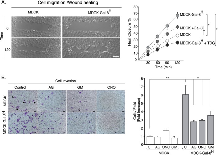
FIGURE 4:
Gal-8 increases migration and invasion capabilities of MDCK cells. (A) Wound healing assay. A confluent monolayer of MDCK and MDCK-Gal-8H cells was scraped with a micromanipulator and pictured every 30 min for 2 h. The picture depicts wound healing at 120 min, while the graph illustrates healing progression under the indicated conditions, including the effects of TDG (20 mM) and exogenous Gal-8 (50 μg/ml) (n = 6 wounds from three experiments). Mean ± SEM; *p < 0.05; two-way ANOVA followed by Sidak’s multiple comparisons test; Scale bar = 40 μm. (B) Invasion assay. MDCK and MDCK-Gal-8H cells (5 × 10⁴) were seeded in Transwell filters (8-μm pore) coated with Matrigel and incubated in the absence or presence of AG1478, GM6001, and ONO4817 for 24 h. Cells stained with crystal violet (arrowheads) were counted on bottom sides of the filter. Graph shows number of cells per field (n = 6 fields from three experiments) Mean ± SEM; *p < 0.05; **p < 0.005; one-way ANOVA, Tukey’s multiple comparisons test).

