FIGURE 6:
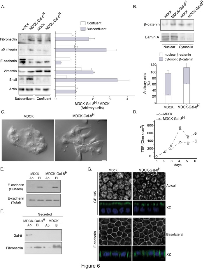
Gal-8 promotes reversible EMT maintaining the capability to generate transepithelial resistance and apical (Ap)/basolateral (Bl) polarity. (A) Subconfluent MDCK-Gal-8H cells display hallmarks of EMT compared with MDCK cells, such as increased levels of fibronectin, α5-integrin, vimentin, and Snail, accompanied by lower levels of E-cadherin. These changes are reverted in confluent cells. Graph shows the mean ratio of MDCK-Gal-8H vs. MDCK cells in arbitrary units of each band density relative to actin in subconfluent and confluent cells (n = three experiments). (B) Higher nuclear distribution of β-catenin in subconfluent MDCK-Gal-8H cells compared with MDCK cells. Lamin A is used as a marker of nuclear fraction. Graph shows the average percentage of distribution (n = 3 experiments); (C) Subconfluent MDCK-Gal-8H cells show extensions opposing the mass of cell clusters compared with MDCK cells, without acquiring a complete mesenchymal phenotype; Scale bar = 10 μm. (D) MDCK-Gal8H grown to confluence on Transwell filters (0.4-μm pore) develop more rapidly and higher levels of TER than MDCK cells. (E) Domain-selective biotinylation shows similar basolateral distribution and cell-surface levels of E-cadherin in Transwell-seeded MDCK-Gal8H and MDCK cells. Total E-cadherin corresponds to 5% of cell extract (50 μg/protein). (F) Immunoblot in 18 h conditioned media shows apical secretion of Gal-8 and basolateral secretion of fibronectin. (G) Apical GP135 and basolateral E-cadherin immunofluorescence analyzed in a Leica SP8 scanning confocal microscope. Images of apical, basolateral, and X-Z sections are displayed. GP135 displays a clustered pattern in MDCK-Gal8H cells. Scale bar = 10 µm.

