FIGURE 1:
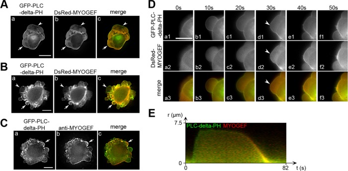
MYOGEF is localized to the bleb membrane during bleb retraction. (A, B) The localization of MYOGEF to the bleb membrane in MDA-MB-231 (A) and M2 melanoma (B) cells. Note that DsRed-MYOGEF was colocalized with the membrane marker GFP-PLC-delta-PH at the bleb membrane in some of the blebs (arrows), but not in others (arrowheads). Bar, 10 μm. (C) The localization of endogenous MYOGEF at the bleb membrane. M2 melanoma cells exogenously expressing GFP-PLC-delta-PH were subjected to immunofluorescence staining for endogenous MYOGEF with a mouse monoclonal antibody (mAb) specific for MYOGEF. Note that endogenous MYOGEF was localized to the bleb membrane (arrows). (D) Time-lapse images showing the localization of MYOGEF at the retracting bleb membrane. DsRed-MYOGEF was recruited to the bleb membrane when the bleb started retracting (arrowheads). Bar, 10 μm. (E) Kymograph showing the localization of DsRed-MYOGEF and GFP-PLC-delta-PH in a bleb cycle. Green indicates the localization of GFP-PLC-delta-PH alone to the expanding bleb membrane, while yellow indicates the colocalization of DsRed-MYOGEF and GFP-PLC-delta-PH to the retracting bleb membrane.

