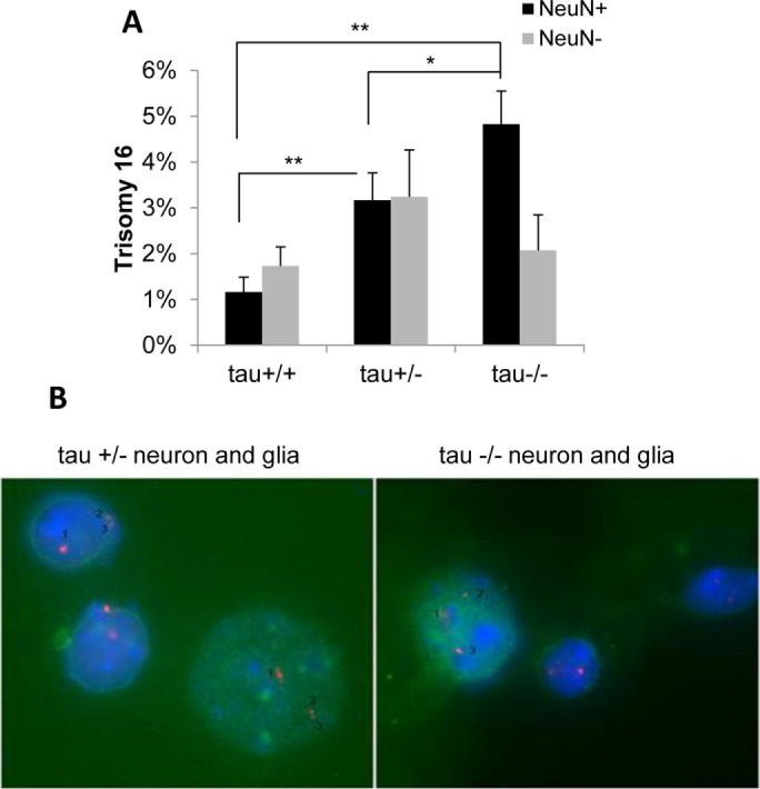
FIGURE 3:

Complete or partial loss of tau function induces trisomy 16 in mouse neurons. Single-cell suspensions of brain cells from tau+/+, tau+/–, and tau–/– mice were analyzed by FISH using a mouse chromosome 16 BAC DNA probe and costained with the anti-NeuN antibody to identify neurons. Quantitative analysis of BAC signals (Spectrum Orange d-UTPs) revealed a significant increase in trisomy 16 in neurons from tau+/– and tau–/– mice compared with tau+/+ mice (A). The graph shows mean difference between groups using ANOVA with Tukey HSD post hoc (effect size η2 = 0.75 for neurons). Representative examples of tau+/– and tau–/– neurons [NeuN(+), green] and glia [NeuN(–)] are shown (B). *p < 0.05, **p < 0.005.
