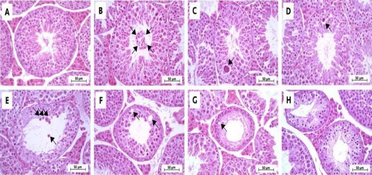
Figure 6.
Representative photomicrographs showing normal histology of the control group (A) and histopathology of seminiferous tubules found in the MLD-STZ group (B-H). Normal arrangement of spermatogenic and Sertoli cells (A), sloughing of deciduous and spermatogenic cells (arrows) into the tubular lumen , large-sized nucleus or bi-nucleated cells (arrow) in the seminiferous epithelium (C), small-sized nucleus cells with vacuolization (arrow) in the seminiferous epithelium (D), few layers of spermatogenic cells and artifactual sloughing (arrows) of germ cell elements into the lumen (E), vacuolation (arrows) of Sertoli cell and absence the spermatids (F), atrophy with germ cell degeneration and small vacuolization (arrow) between spermatogonia and Sertoli cells (G), and the hypospermatogenesis with all germ layers diminished (H

