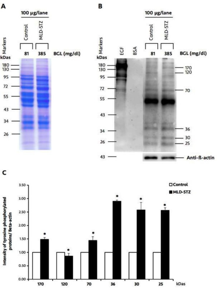
Figure 8.
Representative testicular protein profile revealed by SDS-PAGE (A), immuno-Western blotting of testicular phosphorylated proteins, and relative intensity of testicular phosphorylated proteins (170, 120, 70, 36, 30, and 25 kDas; C) compared between the control and MLD-STZ groups. Epidermal growth factor (EGF)-like factor and bovine serum albumin (BSA) used as positive and negative controls for phosphotyrosine antibody. β-actin was used as an internal control. BGL: blood glucose levels

