Figure 1.
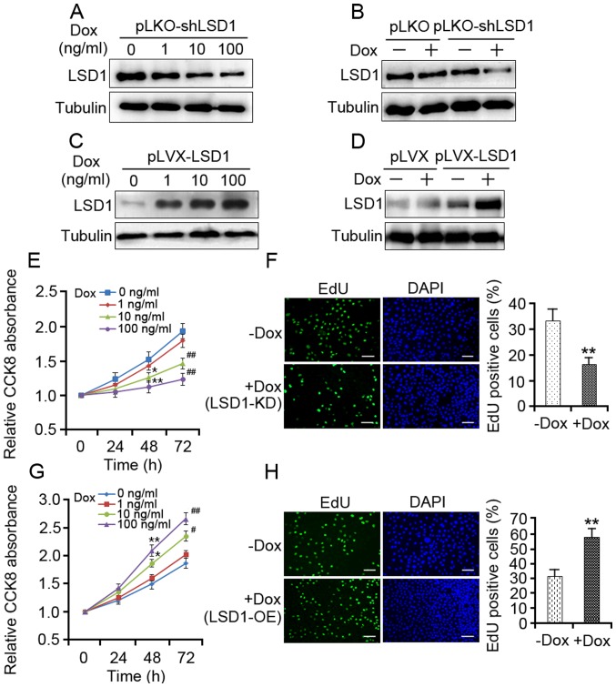
LSD1 is required for the proliferation of SKOV3 cells. (A) SKOV3 cells transduced with pLKO-LSD1-shRNA lentivirus were treated with different dosages of Dox for 48 h. LSD1-KD was determined by western blotting. (B) pLKO and pLKO-LSD1-shRNA-transduced cells were treated with 100 ng/ml Dox for 48 h followed by western blot analysis. (C) LSD1-OE SKOV3 cells were incubated with doses of Dox for 48 h, as indicated; the protein expression levels of LSD1 were assessed by western blotting. (D) pLVX and pLVX-LSD1-transduced cells were treated with 100 ng/ml Dox for 48 h. Subsequently, the protein expression levels of LSD1 were detected via western blotting. (E) LSD1-KD cells were treated with doses of Dox as indicated, and cell viability was assessed using the CCK-8 assay at the indicated durations. (F) LSD1-KD cells were treated with 100 ng/ml Dox for 48 h, and cell proliferation was assessed using the EdU incorporation assay (green). (G) LSD1-OE cells were treated with doses of Dox as indicated, and cell viability was assessed using the CCK-8 assay at the indicated durations. (H) LSD1-OE cells were treated with 100 ng/ml Dox for 48 h, and cell proliferation was assessed using the EdU incorporation assay. Cells of the new generation were detected via EdU (green). DAPI stained nuclei (blue). Error bars represented the data as the mean ± standard deviation (E and G, n=3; F and H, n=4). *P<0.05 and **P<0.01, compared with the group not treated with Dox (48 h); #P<0.05 and ##P<0.01, compared with the group not treated with Dox (72 h). Scale bar=50 µm. CCK-8, Cell Counting Kit-8; Dox, doxycycline; EdU, 5-ethynyl-2′-deoxyuridene; LSD1, lysine-specific demethylase 1; shRNA, short hairpin RNA; pLKO, empty vector; pLVX, empty vector; LSD1-KD, Dox-mediated LSD1 knockdown of cells transduced with pLKO-LSD1-shRNA; LSD1-OE, transduced with lentivirus expressing pLVX-LSD1.

