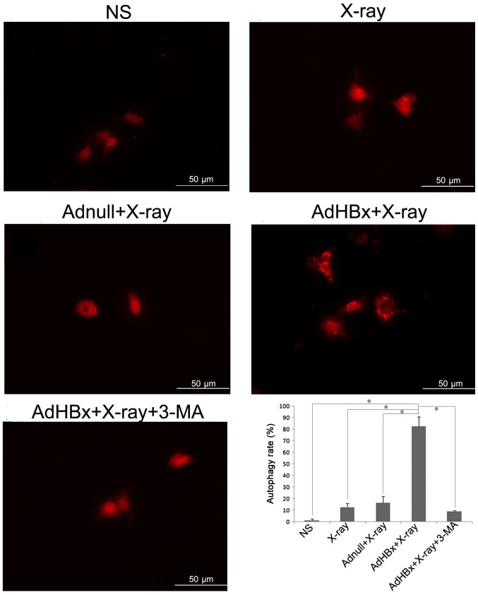
Figure 1.
Autophagy measured by changes in GFP-LC3 puncta. Hepa1-6 cells were transfected with GFP-LC3 prior to each treatment, then cells were fixed and examined by confocal microscopy for changes in GFP-LC3 puncta. Autophagy rate was also examined under 10× magnitude. In normal condition, RFP-LC3 fusion protein diffuse in the cytoplasm (LC3-I); when autophagy occurs, RFP-LC3 fusion protein (conversion of LC3-I to LC3-II) translocate to the autophagosome membrane, forming bright red fluorescent autophagosomes (puncta) (irradiated AdHBx-infected Hepa1-6 cells) under the fluorescence microscope which was reflected by significantly high levels of puncta in cells; *P<0.05 as indicated.

