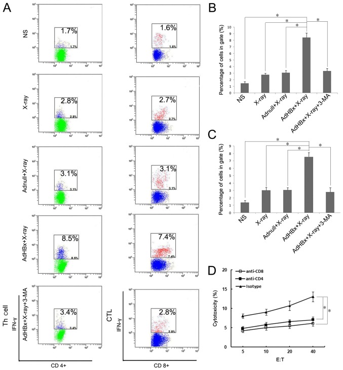
Figure 5.
Detection of specific T responses after priming with DCs. Naïve T-cells were isolated from spleens of C57BL/6 mice and co-cultured with DCs pulsed by NS, irradiated Adnull-infected Hepa1-6 cells, irradiated Hepa1-6 cells, irradiated AdHBx-infected Hepa1-6 cells or irradiated AdHBx-infected Hepa1-6 cells co-treated with 3-MA. The Golgi Stop was added during the last 4–6 h. The cells were then harvested and stained by anti-CD4-APC and anti-IFN-γ-PE or anti-CD8-FITC and anti-IFN-γ-PE. Fluorescence profiles were acquired on a FACScan and analyzed using FlowJo software version 7.6.1 (A) Percentage of the IFN-γ expressing CD4+ T cells (B) and IFN-γ expressing CD8+ T cells (C) were shown as mean ± SEM (*P<0.05). To determine the role of CD4+ or CD8+ T cells in antitumor effect, anti-CD8 or anti-CD4 antibodies were used to deplete corresponding T-cell subsets one day before co-incubation with pulsed DCs, lymphocytes stimulated with pulsed DCs were added to fresh CFSE-labeled target cells at various E:T ratios for 4 h at 37°C. Following incubation, PI was added to each sample. Then, specific cytotoxic activity was performed using flow cytometric analysis and the percentage of CFSE+PI+ cells was considered as specific T cell cytotoxic lysis when the E:T ratios ≥5; *P<0.05. Depletion of either CD8+ T cells or CD4+ T cells during priming almost abrogated the cytotoxicity against Hepa1-6 (D).

