Figure 3.
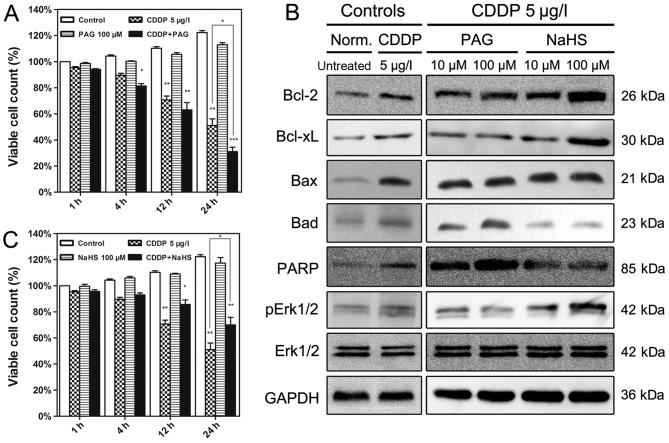
Effects of PAG and NaHS on cell viability and the expression of H2S-associated proteins in EJ cells. (A) EJ cells received different treatments for different time periods. The cells treated with 100 µM PAG were the most affected by treatment with CDDP. (B) Western blotting was employed to verify the modulation of apoptosis-associated protein expression at 12 h. (C) NaHS (100 µM) significantly alleviated the cytotoxicity of CDDP in EJ cells at different time periods. Data are presented as the means ± standard deviation of three experimental repeats. *P<0.05, **P<0.01, ***P<0.001 vs. Control. PAG, propargylglycine; CDDP, cisplatin; Bcl-2, B-cell lymphoma 2; Bcl-xL, Bcl-2-like 1; Bax, Bcl-2-associated X; Bad, Bcl-2-associated agonist of cell death; PARP, poly(ADP-ribose) polymerase; Erk1/2, extracellular-signal-regulated kinase 1/2; pErk1/2, phosphorylated Erk1/2; Norm., normal.

