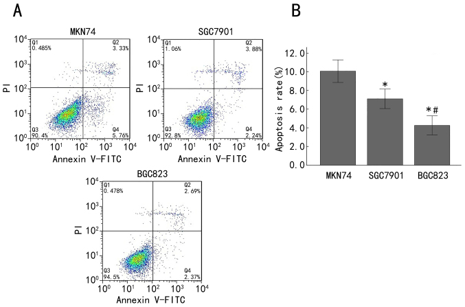
Figure 4.
Apoptosis was increased in late-stage gastric adenocarcinoma cell lines. (A) Apoptosis was analyzed using Annexin V-FITC/PI staining and flow cytometry in MKN74, SGC7901 and BGC823 cells. The right lower quadrant demonstrates early apoptotic cells and the right upper quadrant demonstrates late apoptotic cells. (B) Quantification of the percentage of apoptotic cells in MKN74, SGC7901 and BGC823 cells. *P<0.05 compared with the MKN74 cells; #P<0.05 compared with the SGC7901 cells. FITC, fluorescein isothiocyanate; PI, propidium iodide.

