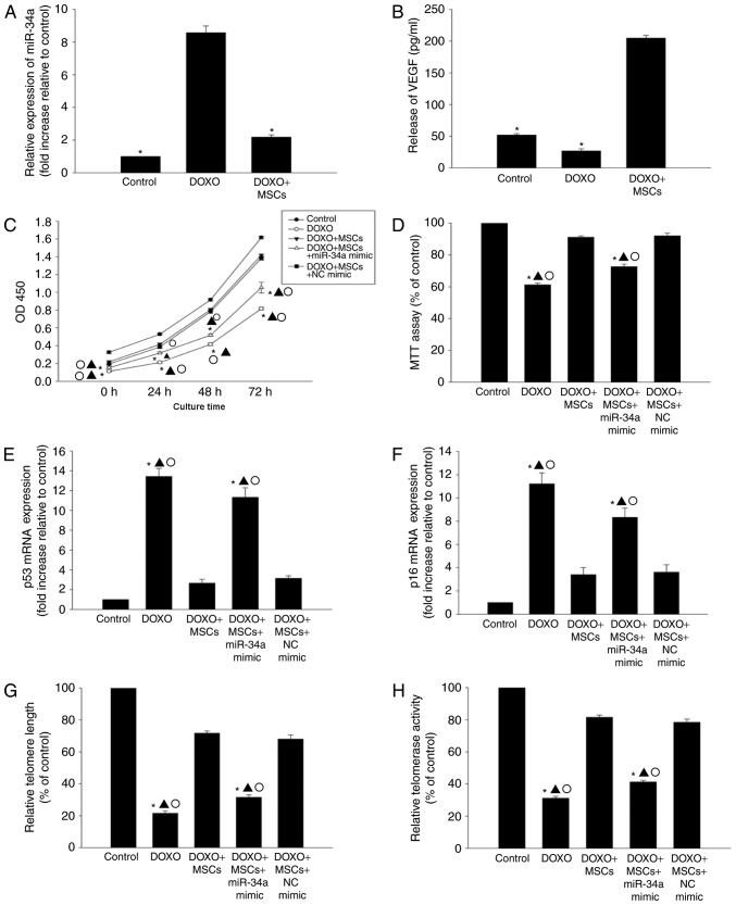
Figure 3.
MSCs induce anti-senescence by inhibiting miR-34a expression. (A) RT-qPCR analysis of the levels of miR-34a mRNA in H9c2 cells incubated with DOXO or co-cultured with MSCs in the presence of DOXO. Each data point represents the mean ± standard deviation of three independent experiments. *P<0.05 vs. DOXO. (B) ELISA analysis of the levels of VEGF released from H9c2 cells that were incubated with DOXO or co-cultured with MSCs. Each data point represents the mean ± standard deviation of three independent experiments. *P<0.05 vs. DOXO+MSCs. (C) Proliferation was determined using Cell Counting Kit-8 assay in several treatment groups: i) DOXO-treated H9c2 cells; ii) co-culture with MSCs and treatment with DOXO; iii) co-culture with MSCs and transfection with a miR-34a mimic in the presence of DOXO and iv) co-culture with MSCs and transfection with NC mimic in the presence of DOXO. (D) The viability of H9c2 cells was analyzed using MTT assay. The analysis of (E) p53, (F) p16 mRNA levels and (G) telomere length was performed using RT-qPCR. (H) The relative telomerase activity was measured. Each data point represents the mean ± standard deviationof three independent experiments.*P<0.05 vs. control; ▲P<0.05 vs. DOXO+MSCs; ○P<0.05 vs. DOXO+MSCs+NC mimic. MSCs, mesenchymal stem cells; miR, microRNA; RT-qPCR, reverse transcription-quantitative polymerase chain reaction; DOXO, doxorubicin; VEGF, vascular endothelial growth factor.

