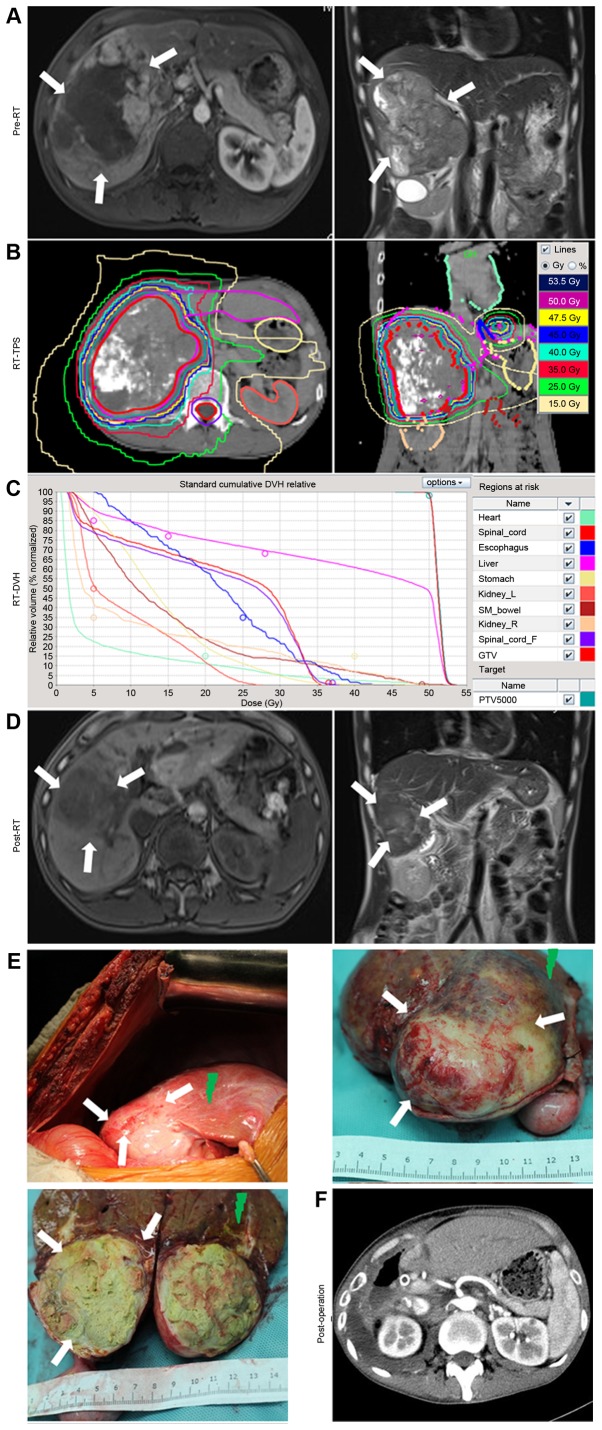
Figure 1.
Example of the treatment of a patient with locally advanced hepatocellular carcinoma using RT followed by resective surgery. (A) Pre-RT MRI revealed a large mass signal on the enhanced T1-W1. (B) Isodose distribution graphs for RT, as produced by the RT-TPS computers. The tumor, liver, stomach, esophagus, heart, kidneys and spinal cord were delineated. (C) RT-DVH graph of the tumor, liver, stomach, esophagus, heart, kidneys and spinal cord, as calculated using the TPS. (D) Pre-operative MRIs revealed that the tumor had regressed post-RT, and was now suited for further treatment with surgery. (E) During surgery, it was identified that the remaining tumor had been almost eliminated by RT, with a distinct boundary (white arrows). Radiation-induced liver diseases occurred in the swelling and bleeding zone of the peritumoral liver tissues (green arrows). (F) Computed tomography scans revealed the absence of the right liver lobe and tumor tissue following surgery. RT, radiotherapy; MRI, magnetic resonance imaging; TPS, treatment planning system; DVH, dose volume histogram.

