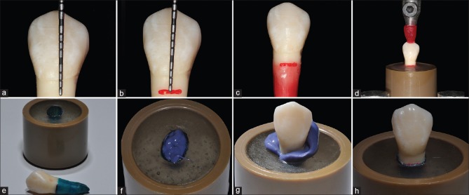
Figure 1.
Periodontal ligament simulation steps and teeth embedded in acrylic. (a and b) Measuring (a) and marking (b) 2 mm below the cement-enamel junction. (c) 0.3 mm wax layer. (d) Inclusion in acrylic resin. (e) Recovering the roots with polyether adhesive. (f) Inserting of the impression material. (g) Tooth placement in the cylindrical device. (h) Periodontal ligament simulation finalized

