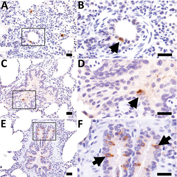
Figure 4.

Influenza D virus immunohistochemistry in swine lung at 3 days (A and B), 5 days (C and D), and 7 days (E and F) postinoculation. Right column panels are higher magnification of boxed region in panels to the left. At all time points, scattered immunopositive bronchiolar epithelial cells were observed (arrows). Scale bars indicate 20 µm.
