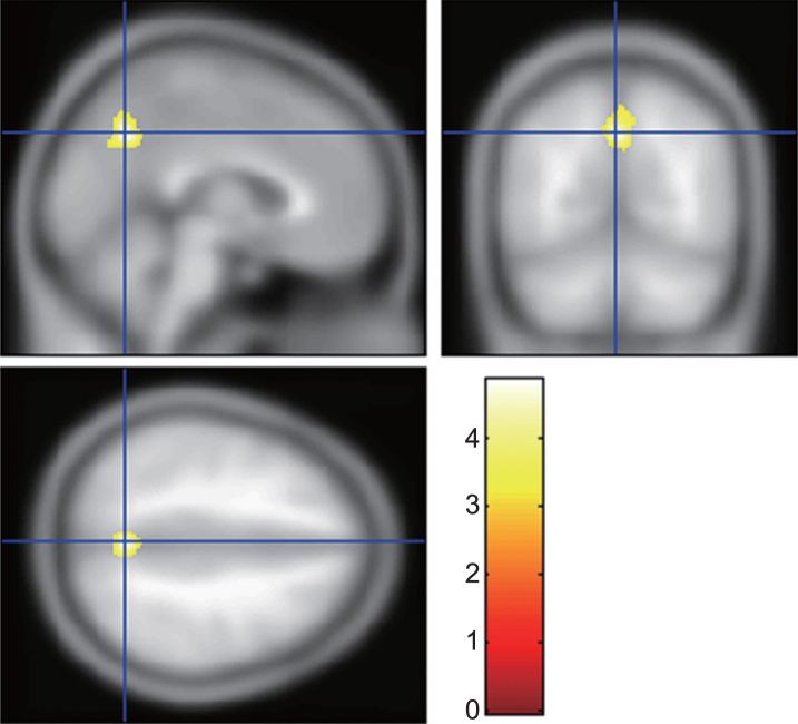
Fig.2.
Brain region involved in sleep disturbance in Alzheimer’s disease (AD). The gray matter volume of the precuneus in AD with sleep disturbance was significantly smaller than that in AD without sleep disturbance. The statistical thresholds were set to uncorrected p-values of 0.001 at the voxel level and to corrected p-values of 0.05 at the cluster level.

