Figure 1.
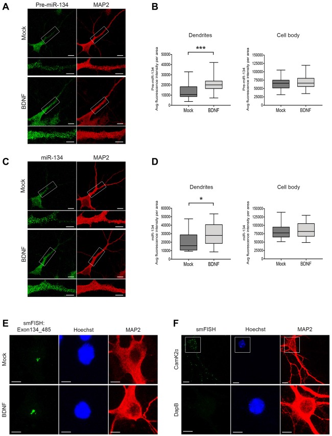
Short-time BDNF treatment induces pre-miR-134 dendritic accumulation. (A,C) Representative pictures from fluorescence in situ hybridization (FISH) using probes directed against the pre-miR-134 loop (A) or mature miR-134 (C) performed in developing rat hippocampal neurons (DIV7) treated with BDNF for 2 h (scale bar: 10 μm). Inserts at higher magnification illustrate the dendritic accumulation of pre-miR-134 granules. MAP2 staining (red) was used to visualize dendritic processes (scale bar: 5 μm). (B,D) Quantification of pre-miR-134 (B) or mature miR-134 (D) FISH signal in dendrites (left) or cell bodies (right) of multiple neurons indicated in (A,C). (B) Mock n = 27, BDNF n = 27. (D) Mock n = 27, BDNF n = 28 cells analyzed from n = 3 independent biological replicates. Mann-Witney test, *P < 0.05, ***P < 0.001. (E,F) BDNF-induced increase in pri-miR-134 is confined to the nucleus. (E) Representative pictures of single molecule FISH (smFISH) with probes spanning Exon134_485 of the Mirg transcript. Pictures show a magnified region of the neuronal cell body, no signal was detected in dendrites (scale bar: 5 μm). (F) Representative pictures of smFISH with probes specific for Camk2α (positive control for dendritic RNA localization, scale bar: 10 μm; top) or DapB (bacterially expressed gene used as a negative control, scale bar: 5 μm; bottom).

