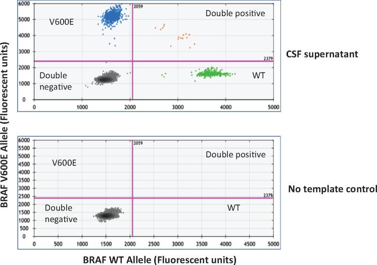
FIGURE 1.
Mutation detection in CSF-ctDNA by ddPCR. Representative 2D plots of ddPCR droplet reads. Bottom plot represents droplet reads from a no-template control sample. All droplets are double negative in the no-template control. The top panel represents the droplet reads from the CSF-cfDNA from Patient 4. Note the presence of WT-DNA in the X-axis (green dots) and BRAF p.V600E mutant DNA in the Y-axis (blue dots). A few droplets contained both WT and mutant DNA and fell in the double positive category (orange dots).

