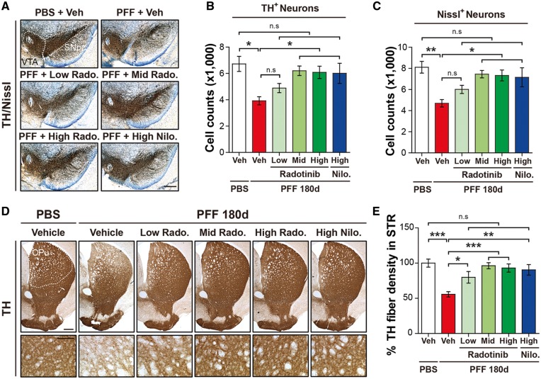
Figure 5.
Radotinib HCl protects against α-synuclein PFF-induced dopaminergic neurodegeneration and fiber density. (A) Representative photomicrographs from coronal mesencephalon sections containing TH-positive neurons in PBS and α-synuclein PFF stereotaxic injected mice treated with vehicle or Radotinib HCl and Nilotinib. The scale bar represents 500 μm. (B) Stereology counts of TH and (C) Nissl-positive neurons in the SNpc region. Unbiased sterologic counting was performed in SNpc region. Error bars represent the mean ± S.E.M, n = 4–6 mice per groups. (D) Representative photomicrograph of striatal sections stained for TH immunoreactivity. High power view of TH fiber density in the striatum (lower panels). The scale bars represent 100 μm (upper panles) and 50 μm (lower panels). CPu and STR, Striatum. (E) Quantification of dopaminergic nerve fiber densities in the striatum by using Image J software (NIH). Error bars represent the mean ± S.E.M, n = 4–6 mice per groups. One-way ANOVA was used to test for the statistical analysis, followed by Tukey’s correction; *P < 0.05, **P < 0.01, ***P < 0.001, n.s., not significant. Radotinib, Radotinib HCl. Nilo., Nilotinib.

