Figure 6.
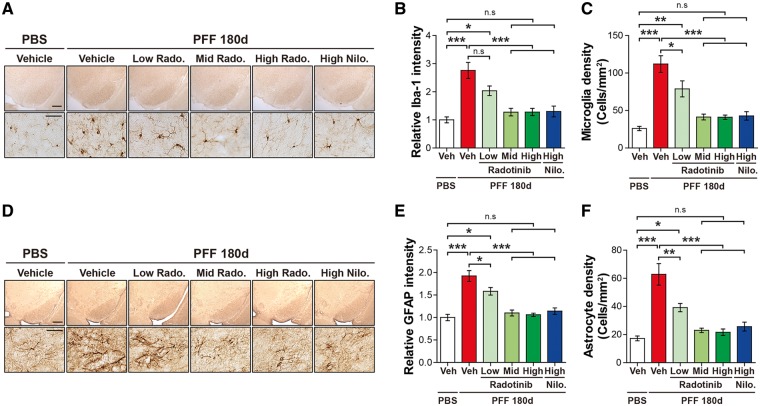
Radotinib HCl restores α-synuclein PFF-induced neuroinflammation. (A) Representative images of immunohistochemistry data for Iba-1 in PBS and α-synuclein PFF stereotaxic injected mice treated with vehicle or Radotinib HCl and Nilotinib. The scale bars represent 500 μm (upper panels) and 50 μm (lower panels). (B) The intensity of Iba-1 positive signals in the SNpc and (C) quantification of Iba-1 positive cell number in SNpc. The intensity of Iba-1 immunoreactivity and densities of microglia in the SNpc region were measured with ImageJ software. Error bars represent the mean ± S.E.M, n = 4–6 mice per groups. (D) Representative images of immunohistochemistry data for GFAP in PBS and α-synuclein PFF stereotaxic injected mice treated with vehicle or Radotinib HCl and Nilotinib. The scale bars represent 500 μm (upper panles) and 50 μm (lower panels). (E) The intensity of GFAP positive signals in the SNpc and (F) quantification of GFAP positive cell number in SNpc. The intensity of GFAP immunoreactivity and densities of astrocyte in the SNpc region were measured with ImageJ software. Error bars represent the mean ± S.E.M, n = 4–6 mice per groups. One-way ANOVA was used to test for the statistical analysis, followed by by Tukey’s correction; *P < 0.05, **P < 0.01, ***P < 0.001, n.s., not significant. Radotinib, Radotinib HCl. Nilo., Nilotinib.

