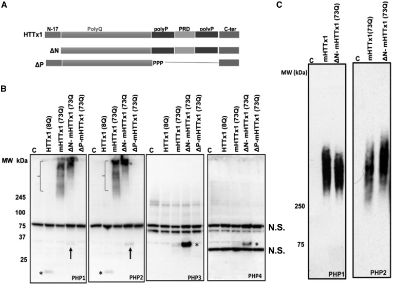
Figure 3.
PHP antibodies bind to HTTx1 assemblies formed in HEK-293 cells. (A) Schematic depiction of constructs used for transfection in part (B). (B) WB analysis of lysates from HEK-293 cells transfected with various constructs of HTTx1. Membranes were probed with PHP1–4 antibodies. Asterisks in the left two panels indicate faint detection of WT HTTx1 (8Q) by PHP1 and PHP2, respectively. Arrows in the left two panels point at weak reactivity of PHP1 and PHP2 to ΔN-mHTTx1. Brackets indicate the assemblies of mHTTx1 and ΔN-mHTTx1 detected by PHP1 and PHP2. PHP3 and PHP4 only detected monomeric mHTTx1 highlighted by asterisks in the right-most two panels, respectively. Part (C) shows examination of HEK-293 cell lysate expressing mHTTx1 and ΔN-mHTTx1 by agarose-SDS gel electrophoresis and probing with PHP1 and PHP2. All experiments were repeated three times. N.S., non-specific bands.

