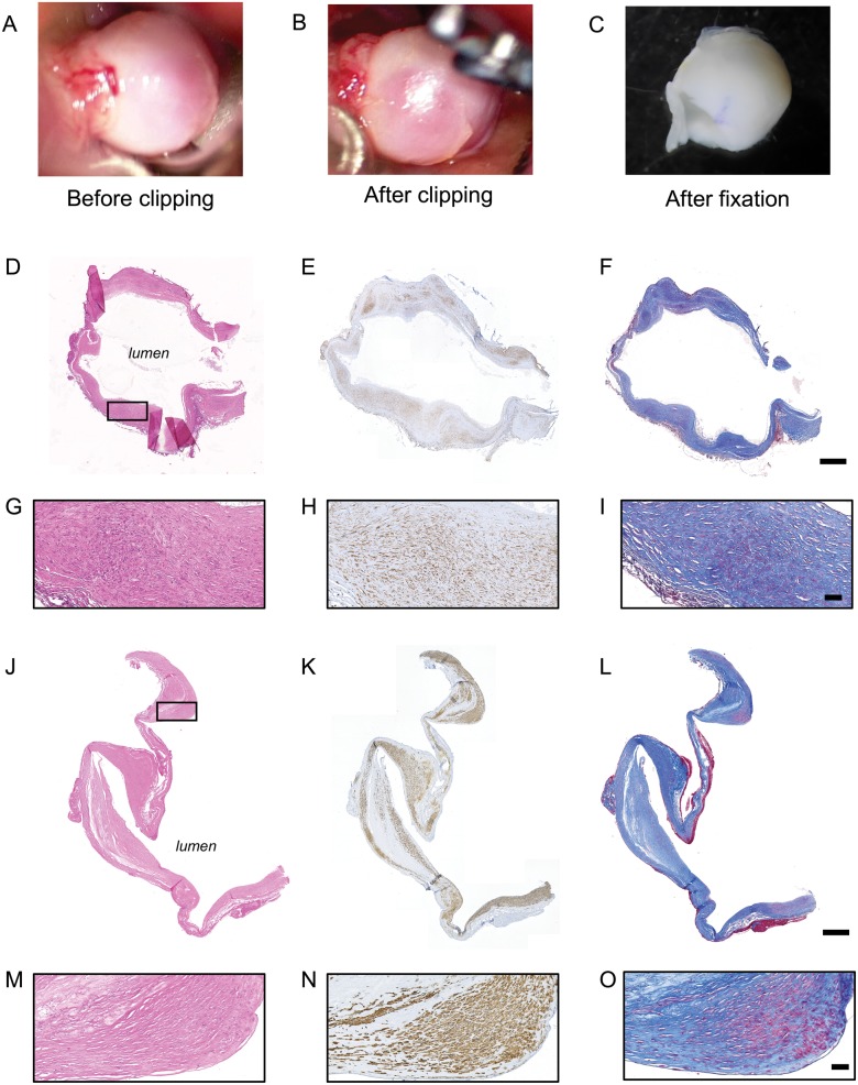
FIGURE 1.
Sample preparation. Examples of intracranial aneurysm (IA) dome obtained during the surgery (before [A] and after [B] the clipping of the IA neck) and just before paraffin embedding (C). Representative examples of H&E (D, G, J, M), α-SMA (SMCs in brown, E, H, K, N) and Masson-trichrome (collagen in blue, F, I, L, O) stainings performed on IA domes stored in RNA latter solution (D–I) or directly fixed in formol (J–O). The black rectangle in panels D and J represent the region of magnification for panels G–I and M–O. Scale bars: D-F, J-L = 500 μm; G-I, M-O = 50 μm.

