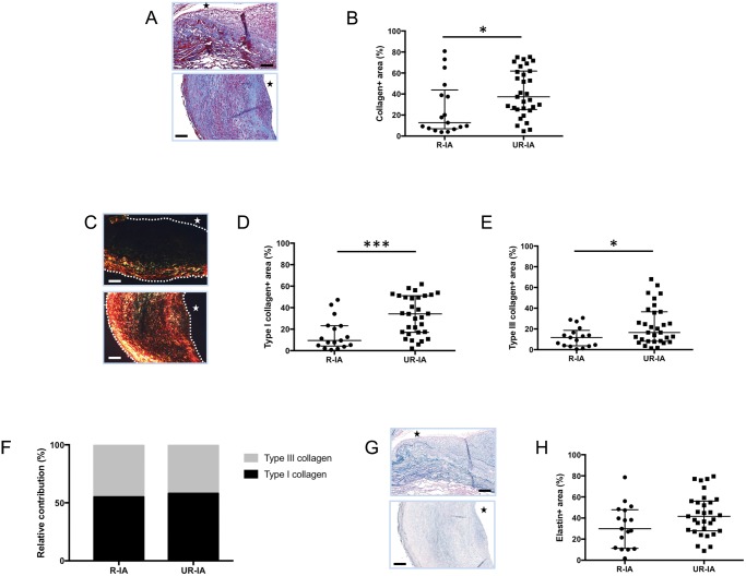
FIGURE 2.
Extracellular matrix composition in ruptured and unruptured intracranial aneurysms (IAs). Representative examples and quantification of total collagen (Masson-trichrome staining: collagen in blue, A, B), type I collagen (Picrosirius red staining: type I collagen in yellow-red, C, D) and type III collagen (Picrosirius red staining: type III collagen in green, C, E) in ruptured (R-IA) and unruptured (UR-IA) IA domes. The relative contribution of type I (black) and type III (grey) collagen is not different between the 2 groups (F). Representative examples and quantification of elastin (Victoria blue staining: elastin in blue, G, H) in ruptured and unruptured IA domes. The lumen of the vessel is indicated by a star. Scale bars in panels A, C, and G represent 100 μm. Dotted lines on the images of panel C represent the borders of the samples. Results are shown as individual values and as median ± interquartile range (B, D, E, and H) or in percentage (F). *p < 0.05, ***p < 0.001, nonparametric Mann-Whitney U test.

