FIGURE 5.
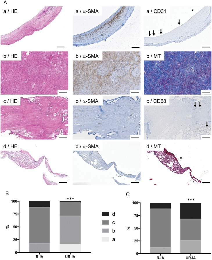
Classification of ruptured and unruptured intracranial aneurysms (IAs) based on histological characteristics. (A) Representative examples of stainings allowing IA histological classification following the definitions: (a) Endothelialized wall with linearly organized SMCs (first row); (b) Thickened wall with disorganized SMC (second row); (c) Hypocellular wall with either intimal hyperplasia or organizing luminal thrombosis (third row), and (d) An extremely thin thrombosis-lined hypocellular wall (fourth row). Scale bars: 100 μm. HE: hematoxylin and eosin staining; α-SMA: α-smooth muscle actin immunostaining (SMCs in brown); CD31: endothelial cell staining (in brown, endothelium indicated by arrows); MT: Masson-trichrome staining (collagen in blue); CD68: macrophage staining (in brown, some macrophages are indicated by arrows). The lumen of the vessel is indicated by a star. (B, C) Classification of ruptured and unruptured IAs following the (a, very light gray), (b, light gray), (c, dark grey) and (d, black) classification based on the dominant wall type (B) or on the most pathological region (C) observed in the resected sample. ***p < 0.001, Chi-square test.

