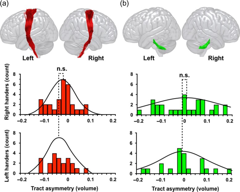
Figure 2.
The distribution of hemispheric asymmetry of the (a) corticospinal tract and (b) superior cerebellar peduncle in right-handers (top row) and left-handers (bottom row). A negative laterality index reflects larger tract volume in the left hemisphere than the right. The tractography images of the corticospinal tract and superior cerebellar peduncle are from a left-hander participant.

