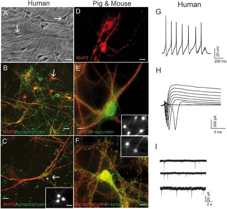
FIGURE 1.
Human, pig, and mouse forebrain neurons in cell culture characterized by immunofluorescence for neuron-specific markers, and human neurons characterized by patch-clamp recording. (A) Phase contrast microscopy of live human neurons generated by culturing and differentiating human NSCs derived from the H9 human embryonic stem cell line. The human neurons are well differentiated and healthy and form a network with elaborate dendrites. (B, C) Immunofluorescence for MAP2 (red) and synaptophysin demonstrates that the cultured human cells are neurons that have differentiated elaborate dendrites (MAP2, red) decorated with a rich network of synaptic boutons (green dots). Some neurons have a pyramidal neuron-like morphology (white arrow, asterisk identifies cell nucleus) and are strongly positive for the cortical neuron marker TRB1 (C, inset). (D, E) Pig forebrain NSC-derived neurons were positive for MAP2 (D) and for MAP2 and β-synuclein (E). Many cells were positive for the cortical neuron marker TRB1 (E inset). (F) Mouse forebrain NSC-derived neuronal cultures were enriched in the neuronal markers synaptophysin and β-synuclein. (G) Electrophysiological properties of human NSC-derived neurons. A current-clamp recording from a human neuron showing action potentials in response to current injection. (H) Whole-cell voltage-clamp recordings showing voltage-dependent sodium and potassium currents. (I) Cultured human neurons showed spontaneous postsynaptic currents. Scale bars: A, B = 25 µm; C, D = 15 µm; E, F = 12 µm; C inset, same for E, F insets = 11 µm.

