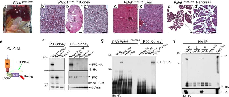
Figure 2. Characterization of Pkhd1Flox67HA mice.
(a) Gross anatomy of a 6-month old Pkhd1Flox67HA mouse showing normal liver and pancreas. Li: liver, Pa: pancreas. (b–d) Representative histologic sections from kidney (b, b’, b”), liver (c, c’) and pancreas (d, d’) derived from a 6-month old Pkhd1Flox67HA mouse. The black dotted lines and squares indicate corresponding enlarged areas. Scale bars represent 1mm in panel b (magnification 1×) and 200µm in panels b’, b”, c, c’, d and d’. Magnification: c, d 4 ×, Magnification b, b’, b”, c’, d’ 10 ×. (e) Schematic representation of the FPC C-terminal transmembrane fragment (FPC PTM) including the transmembrane domain (TM), the ciliary targeting sequence (CTS), the polycystin-2 binding domain (PC2BD) the nuclear localization signal (NLS) and 3 HA tags. The epitope recognized by the mFPC-ct antibody is contained within the bracketed region. (f) Western blot analysis of total lysates prepared from murine kidneys of the indicated genotypes at post-natal day 0 (P0) and 30 (P30). Western blots were probed with antibodies against HA (upper panel), mFPC-ct (middle panel) and actin as a loading control (lower panel). The asterisk indicates a non-specific band in this blot recognized by the mFPC-ct antibody. (g) Western blot survey of total lysates prepared from adult Pkhd1Flox67HA tissues probed with anti-HA. Total lysates from Pkhd1LSL, Pkhd1Δ67 and Pkhd1wt kidneys serve as negative controls. (h) Anti-HA Affinity Matrix was used to Immunoprecipitate (IP) HA tagged FPC from adult kidney (K), liver (L), and pancreas (P) (upper panel). Western blots were prepared and probed with anti-HA antibodies. IP from Pkd1BAC-HA lungs and Pkhd1Δ67 kidneys serve as positive and negative controls, respectively. Overnight exposure of the blot (lower panel) demonstrates liver expression of FPC.

