
Figure 1. Anti-RSV F antibodies isolated from infant B cells display limited SHM and biased V-gene usage.
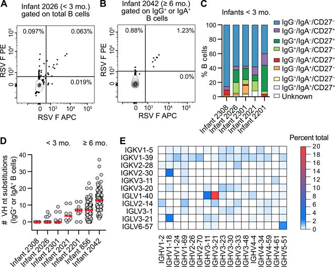
(A) Representative flow plot of the RSV F-specific B cell response in an infant < 3 mo. B cells were gated on CD14− CD8− CD3− CD19+ CD20+ before sorting on double-positive staining with dual-labeled RSV F probes. RSV F-specific B cells are in the upper-right quadrant. Results are representative of at least two independent experiments.
(B) Representative flow plot of the RSV F-specific B cell response in an infant ≥ 6 mo. B cells were gated on CD14− CD8− CD3− CD19+ CD20+ and IgG+ or IgA+ prior to sorting on double-positive staining with dual-labeled RSV F probes. RSV F-specific class-switched B cells are in the upper-right quadrant. Results are representative of at least two independent experiments.
(C) Index sort analysis of surface markers expressed on B cells from which RSV F-reactive antibodies were isolated. Infants are ordered from youngest to oldest at the time of hospitalization, left to right.
(D) Number of VH nucleotide substitutions for antibodies isolated from RSV F-specific class-switched B cells. Infants are ordered from youngest to oldest, left to right. Red bars indicate medians.
(E) Heat map of V-gene usage for all infants. Genes for which no VH:VL pairs were utilized in ≥ 0.5% of antibodies are omitted for clarity.