
Figure 1. Overview of CA1 circuitry.
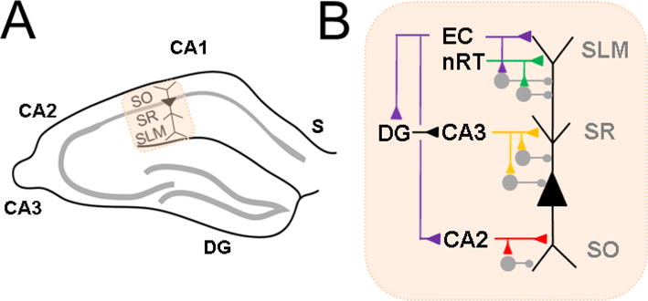
A Hippocampal CA1 in relation to other subregions: subiculum (S), CA2, CA3, dentate gyrus (DG). CA1 pyramidal neuron is indicated with labeled strata stratum oriens (SO), stratum radiatum (SR) and stratuma lacunosum moleculare (SLM). B. CA1 pyramidal neuron excitatory (triangle) and inhibitory (circle) inputs to different parts of its apical dendrite in SR, SLM and basal dendrite in SO. Distal apical dendrite receives direct input from entorhinal cortex (EC) and nucleus reuniens of thalamus (nRT). Indirect EC input arrives at the proximal apical dendrite, via DG and CA3, or at the basal dendrite, from CA2. Each pathway can elicit inhibition in feedforward fashion.