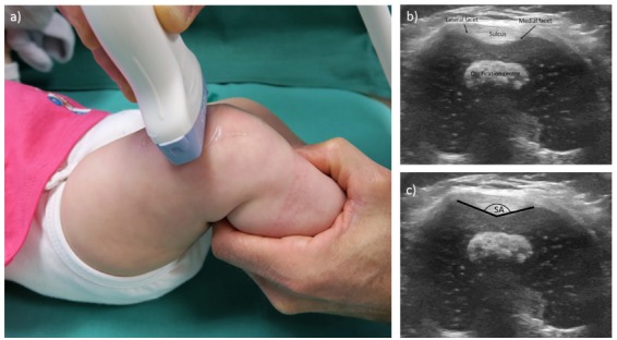
Fig. 1.

Ultrasonographic examination of the femoral trochlea: (a) the ultrasound probe is held perpendicular to the femoral axis levelled at the most ventral point of the lateral facet, obtaining a transverse image of the femoral trochlea (b); (c) the sulcus angle (SA) is shown. The images are produced for illustration of the technique, the child is four months old and not a participant of the study.
