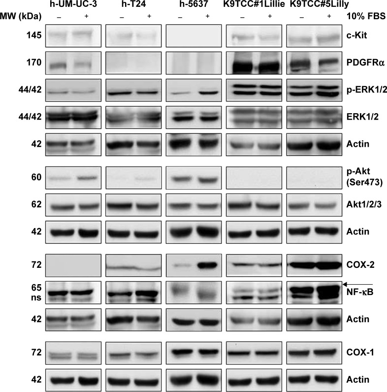
Figure 1.
Expression profile of RTKs and COXs signaling pathway proteins in human and primary canine TCC cells by WB analysis. Cells were grown in the absence (−) or presence (+) of FBS for 24 hours. The expression of c-Kit, PDGFRα, p-ERK1/2, ERK1/2, p-Akt (Ser473), Akt1/2/3, COX-1, COX-2, and NF-κB (specific band labeled with arrow) proteins was evaluated by WB analysis. Actin was used as a loading control. Tested K9TCC cells had higher expression of RTK and COX proteins as compared to h-T24, 5637, and UM-UC-3 cells.
Abbreviations: COX, cyclooxygenase; RTK, receptor tyrosine kinase; WB, Western blot; TCC, transitional cell carcinoma; PDGFR, platelet-derived growth factor receptor; NF-κB, nuclear factor kappa-light-chain-enhance of activated B cells; ERK1/2, extracellular signal regulated kinases 1/2; Akt, V-akt murine thymoma oncogene homolog 1; p-ERK1/2, phosphorylated ERK1/2; p-Akt, phosphorylated Akt; FBS, fetal bovine serum; MW, molecular weight; ns, non-specific.

